
News
Cornea
The cornea is a clear, transparent structure just like our watch glass in the front part of the eye. It plays a key role in vision. As light enters the eye, it is refracted, or bent, by the outside shape of the cornea. The curvature of this outer layer helps determine how well your eye can focus on objects close-up and far away.
There are six main layers of the cornea:
Epithelium- This is the most superficial layer of the cornea. It absorbs oxygen and nutrients from tears.
Stroma- The stroma is the middle and thickest layer of the cornea which is made up mostly of water and proteins that give it an elastic but solid form.
Endothelium- The endothelium is a single layer of cells on the very back of the stroma. The aqueous humor is the clear fluid found in the front chamber of the eye and that is in constant contact with the endothelium. The endothelium works as a pump, expelling excess water back into the eye as it is absorbed into the stroma. Without this specialized function, the stroma would become waterlogged, creating a hazy and opaque cornea and reducing vision.
The term "corneal disease" refers to a variety of conditions that affect mainly the cornea. These include infections, degenerations, dystrophy, trauma, etc.
Cornea can be affected in a variety of disorders and the common symptoms the patient complaint are:
- Pain
- Blurred Vision
- Tearing
- Redness
- Extreme sensitivity to light
Although these symptoms may occur with many other types of eye problems, they may indicate a more serious problem or require special treatment. Therefore, if you experience any of these symptoms, seek care from an eye doctor.
Cornea problems can only be diagnosed after a thorough exam by an eye doctor.
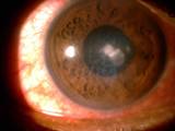
KERATITIS
Keratitis is an inflammation of the cornea that sometimes occurs with infection after viruses, bacteria, or fungi enter the cornea. These microorganisms can enter the eye after superficial or deep injuries, causing infection, inflammation, and ulceration of the cornea. Though uncommon, this type of infection can also arise after injury from wearing contact lenses.
Symptoms of keratitis include:- Severe pain
- Blurred vision
- Tearing
- Redness
- Extreme sensitivity to light
- Discharge
Treatment usually includes antibiotic or antifungal eye drops. Sometimes, antiviral drugs and steroid eye drops are necessary.
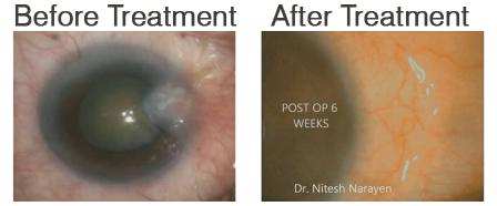
 Before Treatment
 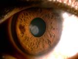 After Treatment
What Is a Pterygium?
A pterygium, from the Greek word for "wing," is an abnormal growth of tissue that extends from the conjunctiva (a membrane that covers the white of the eye) onto the cornea. Pterygia may be small, or grow large enough to interfere with vision and cause irritation. These growths are commonly located on the inner corner of the eye.
Symptoms
- Appearance of a raised pink, white, or red lesion on the eye
- Redness and irritation of the eye
- Foreign body sensation
- Decreased or blurry vision
The symptoms described above may not necessarily mean that you have a pterygium. However, if you experience one or more of these symptoms, contact your ophthalmologist for a complete exam.
Causes
The exact cause of pterygia is not well understood. Long-term exposure to sunlight, especially ultraviolet (UV) rays, wind, and chronic eye irritation from dry, dusty conditions seems to play an important causal role.
Risk Factors
Pterygia occur more often in people who spend a great deal of time outdoors, especially in sunny, windy climates
Tests and Diagnosis
A pterygium can be diagnosed during a routine eye examination. Sometimes photos will be taken to document growth or to measure astigmatism (abnormal curvature) from the pterygium that may contribute to decreased vision. It is important to see an eye doctor, since other more serious conditions can appear that are similar to pterygia.